
상악동 거상술을 동반한 강동구임플란트 식립 과정
상악 어금니 부위 치조골이 얇아 그대로 임플란트를 심기 어려운 경우, 상악동 거상술과 골이식을 동반해 뼈를 보강하고 픽스처를 식립한 실제 임상 과정을 전후 사진과 함께 설명드립니다.
안녕하세요, 강동구 명일동 서울더자연치과 대표원장, 한양대학교병원 외래교수 김민수입니다.
오늘은 상악 어금니 부위의 치조골이 얇아 그대로는 임플란트를 심기 어려웠던 분의 사례를, 상악동 거상술과 골이식을 동반한 강동구임플란트 식립 과정과 함께 설명드리겠습니다.
이 사례는 우측 상악 어금니가 상실된 상태에서, 상악동 아래 치조골 두께가 부족해 상악동 거상술과 골이식으로 뼈를 보강한 뒤 픽스처를 식립한 경우입니다. 치조골이 얇을 때 임플란트를 어떻게 계획하는지 전체 흐름을 이해하시는 데 도움이 되었으면 합니다.
어떤 상태로 내원하셨나요?
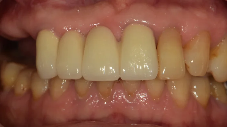
구내 사진을 함께 관찰해보면 우측 어금니가 상실되어 비어 있는 것을 확인할 수 있었습니다.
어금니가 빠진 자리를 오래 비워 두면 저작 기능이 떨어지고, 주변 치아나 맞물리는 치아에도 영향을 줄 수 있습니다.
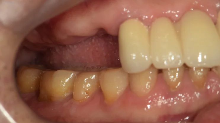
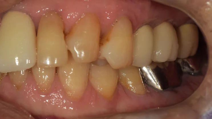
측면에서 보면 우측 어금니 부위가 비어 있는 상태가 더 분명하게 보입니다. 이 공간을 임플란트로 회복하기 위해서는, 먼저 임플란트를 지지할 치조골(잇몸뼈)이 충분한지 확인하는 과정이 필요합니다.
상악동과 치조골, 무엇이 문제였나요?
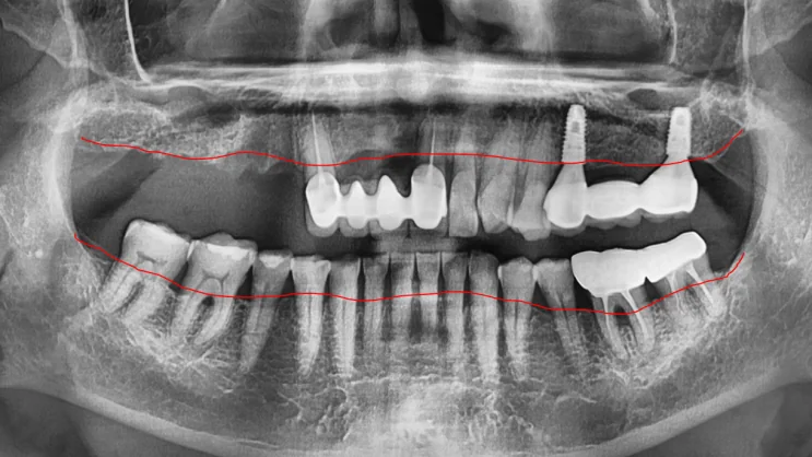
파노라마 사진에서 상악동 아래로 치조골의 두께를 확인해 보니 약 5mm 정도로 판단되었습니다. 이런 경우 9mm 정도의 픽스처를 심기에는 뼈의 양이 부족한 상태에 해당합니다.
상악 어금니 위쪽에는 상악동이라는 빈 공간이 있어, 이 부위는 원래 뼈가 얇은 편입니다.
치조골이 충분하지 않으면 임플란트가 제대로 된 고정력을 얻지 못해 흔들리거나 탈락할 수 있기 때문에, 이런 경우에는 골이식을 동반해 잇몸뼈를 보강해 주는 것이 좋습니다.
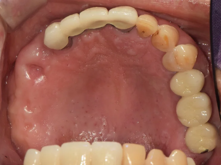
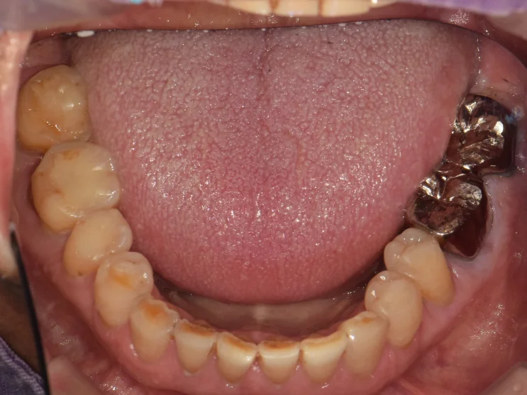
교합면에서 보면 우측 어금니 공간이 비어 있는 것을 확인할 수 있습니다. 이 공간을 채우기 위해 상악동 거상술과 골이식을 동반한 강동구임플란트 식립을 계획했습니다.
상악동 거상술과 골이식을 동반한 식립
상악동 거상술은 상악동 바닥의 점막을 조심스럽게 들어 올린 뒤 그 공간에 뼈를 채워, 임플란트를 지지할 치조골의 높이를 확보하는 방법입니다. 이식해야 할 치조골의 양이 많은 경우에는 측면에서 접근하는 lateral 방식을 사용할 수 있습니다.
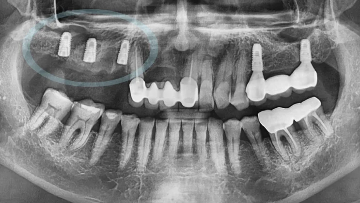
앞서 계획했던 대로 상악동 거상술을 동반하여 픽스처를 식립하고, 엑스레이를 찍어 올바른 위치에 잘 식립되었는지 점검합니다. 이후 임플란트가 단단히 고정될 수 있도록 회복 기간을 가지며, 평균적으로 약 3개월 정도가 소요됩니다.
다만 이식한 뼈의 양이나 골질, 개인 상태에 따라 기간에는 차이가 있을 수 있습니다.
보철 완료와 치료 전후 비교
회복 기간이 지난 후에는 픽스처가 튼튼히 고정되었는지 다시 점검하고, 이후 최종 보철을 수복하면 과정이 마무리됩니다.
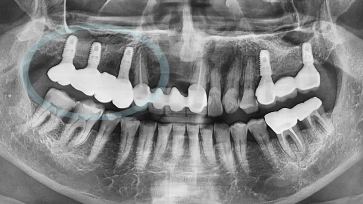
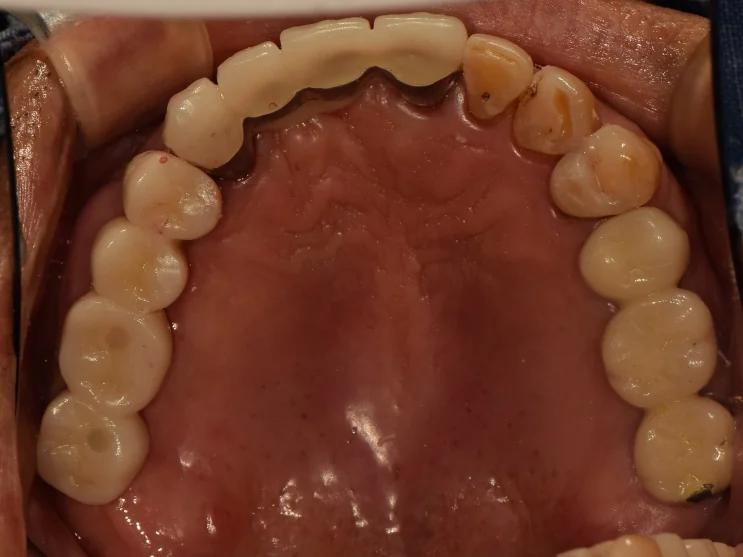
보철 후 모습을 보면 자연치와 유사한 색조와 형태의 지르코니아 보철이 강동구임플란트 어금니 위에 올려져 있는 것으로 보입니다. 비어 있던 우측 어금니 공간이 채워지면서 저작에 사용할 수 있는 치아가 회복되었습니다.
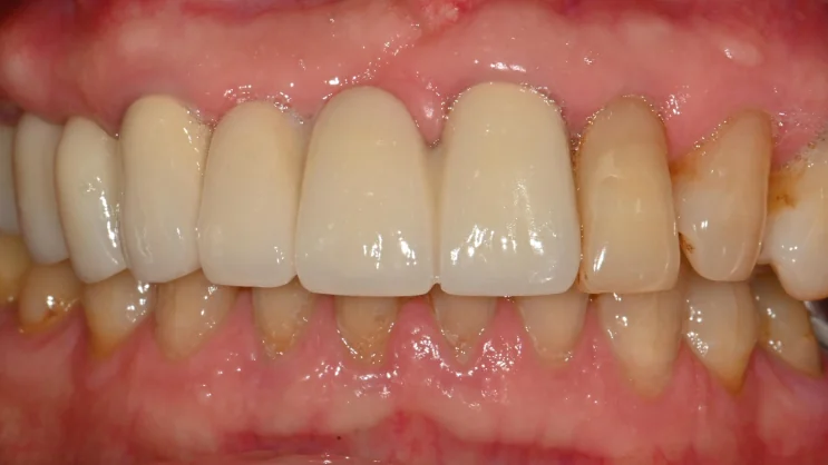
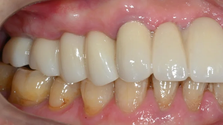
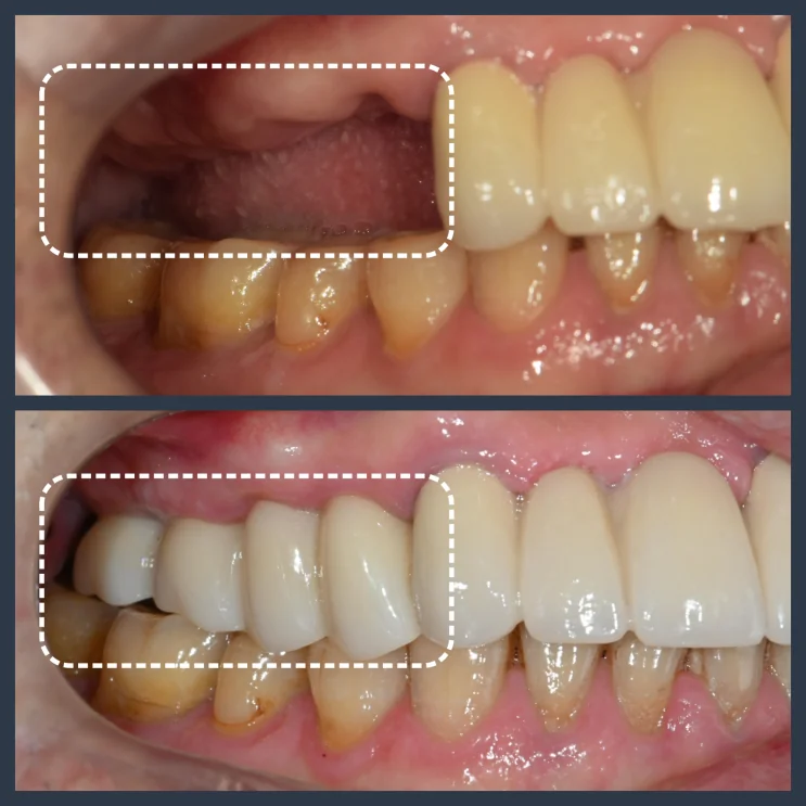
치료 전과 후를 비교해 보면, 비어 있던 우측 어금니 공간이 임플란트 보철로 채워진 변화를 확인할 수 있습니다. 다만 이러한 결과는 진단 시점의 구강 상태에 따른 예시이며, 실제 경과와 결과에는 개인차가 있습니다.
상악동 거상술은 어떤 경우에 필요한가요?
상악 어금니 부위는 상악동이라는 공간 때문에 원래 뼈가 얇은 편이고, 치아가 상실된 뒤 시간이 지나면 치조골이 더 흡수되어 얇아질 수 있습니다. 이렇게 임플란트를 지지할 뼈의 높이가 부족한 경우에 상악동 거상술과 골이식을 함께 고려합니다.
다만 모든 상악 어금니 임플란트에 거상술이 필요한 것은 아니며, 남아 있는 뼈의 높이와 폭, 상악동 상태에 따라 방법과 필요 여부가 달라집니다. 따라서 인터넷의 사례만으로 판단하기보다는, 파노라마나 CT로 치조골과 상악동 상태를 확인한 뒤 계획을 세우는 것이 중요합니다. 강동구임플란트를 알아보실 때도 단순한 개수나 비용보다, 현재 뼈 상태에 맞는 계획이 무엇인지 상담받아 보시길 권해드립니다. 전반적인 내용은 강동구 임플란트 완전 가이드에서도 정리해 두었으니 함께 참고하셔도 좋습니다.
자주 묻는 질문
Q. 상악동 거상술은 왜 필요한가요?
상악 어금니 위쪽에는 상악동이라는 빈 공간이 있어 이 부위는 뼈가 얇은 편입니다. 임플란트를 지지할 치조골의 높이가 부족할 때, 상악동 바닥을 들어 올리고 뼈를 채워 높이를 확보하기 위해 거상술을 고려합니다.
Q. 치조골이 부족하면 임플란트를 못 하나요?
뼈가 부족하다고 해서 반드시 임플란트가 불가능한 것은 아닙니다. 이 사례처럼 상악동 거상술과 골이식으로 뼈를 보강한 뒤 식립하는 방법을 고려할 수 있으며, 부족한 정도와 부위에 따라 계획이 달라집니다.
Q. 골이식한 뼈가 자리 잡는 데 얼마나 걸리나요?
평균적으로 약 3개월 정도의 회복 기간을 두지만, 이식한 뼈의 양과 골질, 전신 상태 등에 따라 차이가 있을 수 있습니다. 회복 후 고정력을 점검한 뒤 최종 보철을 진행합니다.
Q. 상악동 거상술을 동반하면 치료 기간이 더 길어지나요?
뼈를 보강하고 자리 잡는 시간이 필요하기 때문에, 거상술 없이 진행하는 경우보다 전체 기간이 길어질 수 있습니다. 정확한 기간은 뼈 상태와 이식 범위에 따라 달라지므로 검사 후 안내드립니다.
Q. 어금니 공간을 오래 비워 두면 어떻게 되나요?
저작 기능이 떨어질 수 있고, 시간이 지나면서 치조골이 흡수되거나 주변 치아와 맞물리는 치아에 변화가 생길 수 있습니다. 비어 있는 기간이 길수록 뼈 보강이 더 필요해질 수 있어, 변화가 느껴진다면 검진을 받아보시는 것이 좋습니다.
English Summary
Placing an Implant with a Sinus Lift and Bone Graft When Upper Bone Is Thin: A Real Case
This post follows a real clinical case of placing an implant in the upper posterior area when the supporting bone was too thin to accept a fixture directly. The patient had lost a molar on the right upper side, and on examination the ridge beneath the maxillary sinus did not have enough height for a standard implant.
To rebuild that height, a sinus lift together with a bone graft was planned before the fixture was placed, and the empty molar space was later restored with a zirconia crown. The aim was to recover chewing function in an area where the bone alone would not have held an implant securely.
Looking at the intraoral photographs, the right molar area was clearly empty. When a molar space is left unfilled for a long time, chewing efficiency drops and the neighboring and opposing teeth can be affected over time. Before restoring the space with an implant, the first step is to confirm whether there is enough alveolar bone to support a fixture, so a panoramic radiograph was taken to assess the bone height under the sinus.
On the panoramic image, the alveolar bone beneath the maxillary sinus measured only about 5mm in height. For a fixture of around 9mm, that is not enough bone. The upper posterior region naturally has thinner bone because of the maxillary sinus, an air-filled space above the back teeth, and after a tooth is lost the ridge can resorb and become thinner still. When the bone is insufficient, an implant cannot gain proper primary stability and may loosen or fail, so in cases like this a bone graft is added to reinforce the ridge before or during placement.
The plan was therefore to place the implant together with a sinus lift and bone graft. A sinus lift gently raises the membrane at the floor of the maxillary sinus and fills the created space with bone so that enough height is available to support a fixture. When a larger amount of bone needs to be grafted, a lateral approach — accessing the sinus from the side — can be used. Following this plan, the fixture was placed together with the sinus lift, and an X-ray was taken to confirm that it was positioned correctly.
After placement, a healing period is needed so that the graft matures and the implant integrates with the bone. This takes about three months on average, though the amount of grafted bone, the bone quality, and individual factors can change the timeline. Because bone augmentation and healing are involved, the overall treatment period can be longer than a case placed without a sinus lift, which is one reason the schedule is explained individually after examination rather than promised as a fixed number.
Once the healing period had passed, the stability of the fixture was checked again, and the final restoration was then made to complete the process. In the finished photographs, a zirconia crown similar in color and shape to the natural teeth sits on the implant in the previously empty molar area, and the space that had been missing was restored so that the tooth could be used for chewing again. Comparing before and after, the empty right molar space is filled with the implant restoration; that said, this outcome reflects the oral condition at the time of diagnosis, and actual progress and results vary from person to person.
The broader point is that thin upper bone does not necessarily mean an implant is impossible. Not every upper molar implant needs a sinus lift; whether it is needed depends on the remaining bone height and width and the condition of the sinus, which are assessed with a panoramic radiograph or CT before planning.
Rather than deciding from cases seen online, it is better to confirm the bone and sinus condition first and then build a plan suited to the individual. At Seoul The Nature Dental Clinic in Gangdong-gu, Myeongil-dong, patients considering an upper posterior implant are encouraged to focus less on the number of implants or the cost and more on the plan that fits their current bone condition, decided after a proper examination.
마치며
상악 어금니 부위는 상악동 때문에 뼈가 얇은 경우가 많고, 치아를 오래 비워 두면 치조골이 더 흡수될 수 있습니다. 이 사례처럼 뼈가 부족하더라도 상악동 거상술과 골이식으로 잇몸뼈를 보강한 뒤 임플란트를 식립하는 방법을 고려할 수 있습니다.
다만 거상술이 모든 경우에 필요한 것은 아니며, 남은 뼈와 상악동 상태에 따라 방법은 달라집니다. 강동구, 명일동에서 상악 어금니 임플란트나 치조골 부족이 고민되신다면, 무리하지 마시고 먼저 파노라마나 CT 검사를 통해 본인의 뼈 상태에 맞는 계획을 확인해 보시길 권해 드립니다.
지금까지 서울더자연치과 치과의사 김민수였습니다.
※ 본 포스팅은 의료법 제56조 제1항의 의료광고 규정을 준수하여 작성되었으며, 실제 내원 환자분의 동의하에 공개된 사진입니다. 임플란트 및 상악동 거상술·골이식에는 감염, 상악동 관련 합병증, 신경 손상, 보철물 파절 등 부작용이나 주의사항이 있을 수 있고, 개인에 따라 진료 및 치료 방법, 효과와 부작용이 다르게 나타날 수 있으므로, 진료 전 전문 의료진과의 충분한 상담을 권해 드립니다.