
치주염으로 무너진 치아, 발치·골이식·임플란트로 회복한 과정
치주염이 진행되어 뿌리만 남은 치아를 발치하고, 골이식과 함께 임플란트를 식립해 저작 기능을 회복한 실제 과정을 전후 사진과 함께 설명드립니다.
안녕하세요, 강동구 명일동 임플란트 치과 서울더자연치과 대표원장, 한양대학교병원 외래교수 김민수입니다. 오늘은 치주염이 진행되어 여러 치아가 흔들리고 뿌리만 남았던 분의 사례를, 발치와 골이식을 동반한 강동구임플란트 식립 과정과 함께 설명드리겠습니다.
이 사례는 치주염으로 치아를 지지하는 뼈가 손상되어 뿌리만 남은 치아를 발치하고, 인공뼈 이식과 함께 임플란트를 식립한 경우입니다. 치주염이 어떻게 진행되고 어떤 치료로 이어지는지 전체 흐름을 이해하시는 데 도움이 되었으면 합니다.
치주염이란 무엇인가요?
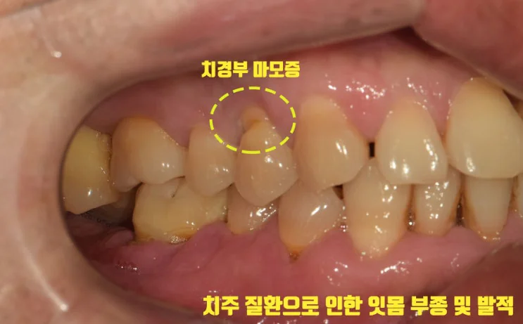
치주염은 치아와 이를 지지하는 조직에 점진적인 손상을 유발할 수 있는 치주 조직의 염증성 질환을 총칭합니다. 흔히 ‘풍치’라고 부르기도 하는데, 잇몸(치은)이 내려앉아 그 아래 덮여 있던 치아 뿌리가 드러나고 이가 흔들리는 데에서 유래한 말입니다.
치은염도 넓게 보면 치주 질환에 속하지만, 둘은 차이가 있습니다. 치은염은 잇몸(치은)에만 한정된 염증을 말하고, 치주염은 잇몸을 넘어 치조골 주변까지 진행된 경우를 말합니다. 치주염 초기에는 잇몸치료만으로도 어느 정도 회복이 가능하지만, 시기를 놓치면 치아를 지지하는 뼈까지 손상될 수 있습니다.
치주염이 진행되면 어떻게 되나요?

초진 파노라마 사진을 관찰해보면, 이미 일부 치아가 뿌리만 남은 상태로 보였습니다. 치주염으로 치아를 지지하는 치조골이 소실되면 치아가 흔들리게 되고, 이 정도로 진행된 경우에는 잇몸치료만으로 회복하기 어려울 수 있습니다.
이가 빠지기 직전 정도로 흔들리는 치아라면 발치가 필요할 수 있고, 이미 치조골 소실이 다소 진행된 경우에는 치조골(골)이식을 함께 계획해 주어야 합니다. 남길 수 있는 치아와 그렇지 않은 치아를 구분하는 것이 치료 계획의 시작입니다.
발치·골이식·임플란트 식립은 어떻게 진행되나요?
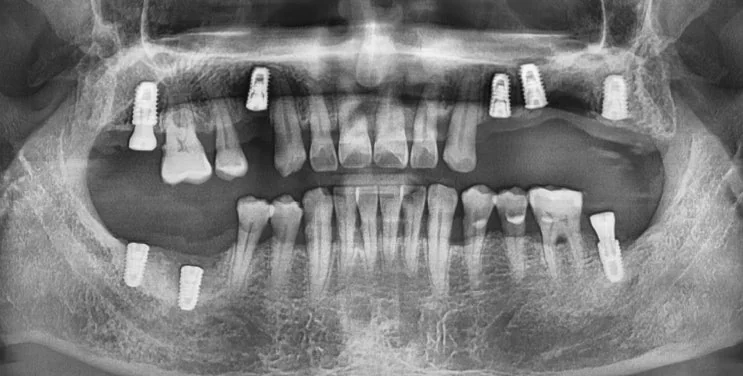
흔들리는 치아를 발치한 뒤, 인공뼈 이식과 함께 임플란트 뿌리(픽스처)를 치조골에 식립합니다. 치주염으로 뼈가 부족해진 부위는 골이식으로 보강해 임플란트를 지지할 수 있는 환경을 만들어 줍니다.
식립 이후에는 치조골과 임플란트 뿌리가 잘 붙기를 기다리는 골 유착 기간이 필요하며, 보통 약 3~6개월 정도 소요됩니다. 이 기간 동안 사용할 임시틀니를 제작해 저작과 심미를 보조할 수 있습니다.
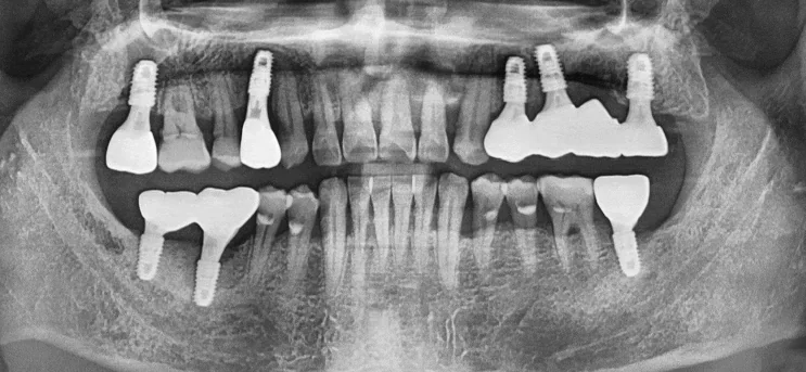
골 유착이 충분히 이루어지면, 식립해 놓은 임플란트 뿌리 위에 크라운을 수복합니다. 다수의 치아가 상실된 경우에도 필요한 위치에 임플란트를 나누어 식립해 저작 기능을 회복할 수 있습니다. 이처럼 여러 치아가 함께 손상된 강동구임플란트 케이스에서는 식립 위치와 개수를 신중하게 계획하는 것이 중요합니다.
명일동 임플란트를 선택한 이유
임플란트는 자연치아와 비슷한 정도의 저작력을 보이고, 안정성이 뛰어나 변형이 적다는 특징이 있습니다. 관리를 잘 하신다면 오래 사용할 수 있습니다.
반면 틀니는 저작력이 자연치아의 40%에 채 미치지 못하는 경우가 많고, 잇몸의 형태가 변하면 금방 잘 맞지 않게 될 수 있습니다. 다만 임플란트와 틀니 중 무엇이 적합한지는 남은 치아와 뼈 상태, 전신 건강, 관리 여건에 따라 달라지므로, 강동구임플란트 상담에서도 검사 후 상담을 통해 결정하는 것이 좋다고 안내드립니다.
치료 전후 비교
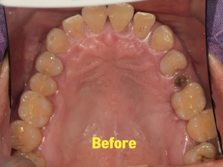
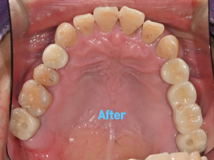
상악 교합면을 치료 전후로 비교해 보면, 치주염으로 무너져 있던 부위가 임플란트 보철로 정리된 변화를 확인할 수 있습니다.

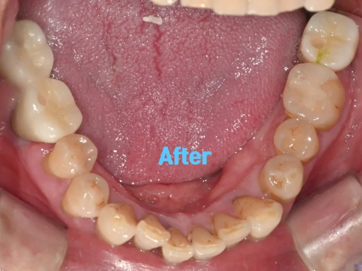
하악 역시 전후를 비교해 보면 저작에 사용할 수 있는 치아가 회복된 모습을 볼 수 있습니다. 다만 이러한 결과는 진단 시점의 구강 상태에 따른 예시이며, 실제 경과와 결과에는 개인차가 있습니다.
강동구에서 치주염과 임플란트를 함께 고민하신다면
치주염은 초기에는 잇몸치료로 관리할 수 있지만, 진행되어 치조골까지 손상되면 발치와 골이식, 명일동 임플란트로 이어질 수 있습니다. 따라서 잇몸 출혈이나 붓기, 치아 흔들림 같은 변화가 있다면 방치하기보다 가능한 이른 시점에 잇몸과 치조골 상태를 확인하는 것이 중요합니다.
이미 뼈 소실이 진행된 경우라도 골이식을 동반한 강동구임플란트로 회복하는 방법을 고려할 수 있으니, 인터넷의 사례만으로 판단하기보다 현재 상태에 맞는 계획을 상담받아 보시길 권해드립니다. 전반적인 내용은 강동구 임플란트 완전 가이드에서도 정리해 두었으니 함께 참고하셔도 좋습니다.
자주 묻는 질문
Q. 치은염과 치주염은 어떻게 다른가요?
치은염은 잇몸(치은)에만 한정된 염증을 말하고, 치주염은 잇몸을 넘어 치조골 주변까지 진행된 경우를 말합니다. 치은염 단계에서는 잇몸치료로 회복 가능성이 높지만, 치주염으로 진행되면 뼈 손상까지 고려해야 합니다.
Q. 치주염이 심하면 임플란트밖에 방법이 없나요?
진행 정도에 따라 다릅니다. 초기에는 잇몸치료로 관리할 수 있고, 뿌리만 남을 정도로 진행되어 보존이 어려운 치아는 발치 후 임플란트나 다른 보철을 고려합니다. 남길 수 있는 치아는 최대한 살리는 방향으로 계획합니다.
Q. 흔들리는 치아는 무조건 빼야 하나요?
흔들리는 정도와 원인, 치조골 상태에 따라 다릅니다. 잇몸치료로 안정되는 경우도 있고, 이가 빠지기 직전으로 흔들린다면 발치가 필요할 수 있습니다. 검사 후 보존 가능 여부를 판단합니다.
Q. 임플란트 골유착 기간은 얼마나 걸리나요?
보통 약 3~6개월 정도가 소요되지만, 골이식 여부와 골질, 식립 부위, 전신 상태 등에 따라 차이가 있을 수 있습니다. 이 기간에는 임시틀니 등으로 불편을 보조할 수 있습니다.
Q. 치주염으로 뼈가 녹았는데 임플란트가 가능한가요?
치조골 소실이 있는 경우에도 골이식으로 뼈를 보강한 뒤 임플란트를 식립하는 방법을 고려할 수 있습니다. 다만 소실 정도와 부위에 따라 계획과 난이도가 달라지므로 검사가 필요합니다.
English Summary
From Advanced Periodontitis to Implants: Extraction, Bone Graft, and Restoration — A Real Case
This post follows a real clinical case in which periodontitis had progressed to the point that several teeth were loose and some were reduced to roots only, and the treatment combined extraction, bone grafting, and implants to restore chewing function. Periodontitis is an inflammatory disease of the periodontal tissues that can cause progressive damage to the teeth and their supporting structures. It is sometimes called “pungchi” in Korean, a term that comes from the way the gum recedes, the previously covered tooth root becomes exposed, and the tooth begins to loosen.
It helps to distinguish gingivitis from periodontitis. Gingivitis is inflammation limited to the gum tissue, whereas periodontitis has progressed beyond the gum to involve the alveolar bone around the tooth. In the early stages, periodontitis can often be managed to some degree with gum treatment, but once the window has passed and the supporting bone is damaged, more involved treatment may be needed. When bone loss has already progressed, a bone graft is planned together with the rest of the treatment so that the ridge can support a restoration.
On the initial panoramic radiograph in this case, some teeth were already reduced to roots only. When periodontitis destroys the alveolar bone that supports a tooth, the tooth loosens, and at that degree of progression gum treatment alone is often not enough to recover it. A tooth that is loose to the point of nearly falling out may need to be extracted, and where alveolar bone loss has advanced, a bone graft is included in the plan. The first step of planning is therefore to separate the teeth that can be saved from those that cannot.
For the treatment itself, the loose teeth were extracted, and an artificial bone graft was placed together with the implant fixtures set into the alveolar bone. Areas where the bone had become deficient from periodontitis were reinforced with the graft to create an environment that could support the implants.
After placement, a period of osseointegration is needed for the bone and the implant to bond, usually taking about three to six months. During this time a temporary denture can be made to assist with chewing and appearance so that daily life is more comfortable while healing proceeds.
Once osseointegration is sufficient, crowns are restored onto the placed implant fixtures. Even when many teeth have been lost, implants can be distributed to the necessary positions to rebuild chewing function rather than replacing every single tooth one for one. The reason implants were chosen over a removable denture in this case is that implants show a chewing force closer to that of natural teeth and are stable with little deformation, so with good maintenance they can be used for a long time.
A removable denture, by contrast, often delivers less than about 40 percent of the chewing force of natural teeth, and when the shape of the gum changes it can quickly stop fitting well. Which option suits a given person still depends on the remaining teeth and bone, general health, and how well the appliance can be maintained, so the choice is made after examination and consultation.
Comparing the before and after photographs of the upper and lower occlusal surfaces, the areas that had broken down from periodontitis were reorganized with implant restorations, and teeth that could be used for chewing were recovered. That said, this outcome reflects the oral condition at the time of diagnosis, and actual progress and results vary from person to person. The broader message is that periodontitis can be managed with gum treatment when caught early, but once it progresses to bone loss it can lead to extraction, bone grafting, and implants. If there are signs such as gum bleeding, swelling, or loosening teeth, it is better to have the gum and alveolar bone assessed as early as possible rather than leaving them.
Even when bone loss has already occurred, restoration with a bone graft and implants may be possible. Because periodontitis can continue to affect the remaining natural teeth, ongoing gum care and regular check-ups after treatment are especially important for keeping both the implants and the natural teeth healthy over time. At Seoul The Nature Dental Clinic in Gangdong-gu, Myeongil-dong, the current condition is examined first so that a plan suited to the individual can be established.
마치며
치주염은 천천히 진행되는 경우가 많아 초기에는 알아차리기 어렵지만, 진행되면 치아를 지지하는 뼈까지 손상되어 발치와 골이식, 임플란트로 이어질 수 있습니다. 이 사례처럼 이미 뼈 소실이 진행된 경우라도 발치와 골이식, 임플란트 식립으로 저작 기능을 회복하는 방법을 고려할 수 있습니다.
다만 치료 방법과 범위는 잇몸과 치조골 상태에 따라 달라집니다. 강동구, 명일동에서 잇몸 출혈이나 치아 흔들림, 치주염과 임플란트가 함께 고민되신다면, 무리하지 마시고 먼저 진료를 통해 본인의 상태에 맞는 계획을 확인해 보시길 권해 드립니다.
지금까지 서울더자연치과 치과의사 김민수였습니다.
※ 본 포스팅은 의료법 제56조 제1항의 의료광고 규정을 준수하여 작성되었으며, 실제 내원 환자분의 동의하에 공개된 사진입니다. 임플란트 및 발치·골이식에는 감염, 신경 손상, 보철물 파절, 골유착 실패 등 부작용이나 주의사항이 있을 수 있고, 개인에 따라 진료 및 치료 방법, 효과와 부작용이 다르게 나타날 수 있으므로, 진료 전 전문 의료진과의 충분한 상담을 권해 드립니다.