
신경관과 가까운 사랑니 발치, 3D CT로 안전하게
사랑니 뿌리끝이 신경관에 닿아 있던 사례입니다. 사랑니 통증의 원인부터, 3D CT로 신경 위치를 확인하고 최소 침습으로 발치한 과정을 사진과 함께 정리해 드립니다.
안녕하세요, 명일동 강동구에 위치한 서울더자연치과 치과의사 김민수입니다. 오늘은 사랑니 뿌리끝이 신경관에 가까이 닿아 있던 사례를, 3D CT로 신경 위치를 확인하며 안전하게 발치한 과정으로 설명드리겠습니다.
사랑니에 통증이 나타나는 이유는 생각보다 여러 가지입니다. 맹출된 형태가 비정상적일 때, 주변 잇몸에 염증이 생겼을 때, 앞 어금니를 짓누르고 있을 때, 사랑니가 많이 썩고 있을 때 등이 대표적입니다. 어떤 이유에서든 통증이나 염증이 생겼다면, 최대한 빠르게 가까운 치과를 방문해 검진을 받아보시는 것이 가장 좋은 방법입니다.
왜 신중한 발치가 필요한 경우였나요?
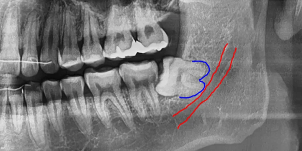
이 사례에서 방사선 사진을 관찰해 보면, 사랑니 뿌리끝이 신경관에 닿아 있는 것으로 판단됩니다. 하치조신경은 아랫입술과 턱 주변의 감각을 담당하는 중요한 신경으로, 이렇게 사랑니 뿌리와 신경관이 가까이 닿아 있는 경우에는 발치할 때 더욱 조심해서 뽑아야 합니다.
신경과 가까운 부위를 무리하게 건드리면 큰 부작용이 생길 수 있기 때문입니다. 따라서 사랑니 뿌리와 신경의 위치를 정확히 파악하는 사전 확인이 안전한 발치의 출발점이 됩니다.
3D CT로 신경 위치를 확인합니다

안전하게 발치하기 위해서는 3차원 CT 촬영을 통해 신경과 사랑니의 뿌리 위치를 정확하게 파악하는 것이 좋습니다. 일반 파노라마는 2차원 영상이라 실제 깊이와 신경과의 관계를 파악하는 데 한계가 있기 때문입니다.
이 사례에서도 CT 촬영본을 통해 사랑니 뿌리와 신경관의 관계를 여러 단면으로 확인했습니다. 거리가 가까운 것은 맞지만, 사전 분석을 통해 발치를 진행할 수 있을 것으로 판단했습니다. 이렇게 신경과 밀접한 경우에는 CT를 통한 사전 확인이 특히 중요합니다.
최소 침습으로 발치를 진행합니다
사랑니를 뺀 후 붓기나 통증이 심하지 않게 발치하기 위해서는 최소한의 조직 침습이 중요합니다. 작게 절개하고 최소한으로 치아 조직을 절제하면 발치 후 통증이 훨씬 적습니다.
그리고 사랑니가 부분 매복되어 있는 경우에는 충치와 치주질환의 발생 확률이 높기 때문에 치아 표면이 부식되어 있을 가능성이 큽니다. 따라서 조각을 낼 때 으스러지지 않도록 신중하게 제거하는 것이 좋습니다. 신경과 가까운 만큼 서두르지 않고 조심스럽게 접근하는 것이 핵심입니다.

발치 후 예시 사진을 보면 잔존 조직 없이 깨끗하게 제거된 사랑니를 확인할 수 있습니다. 신경관에 근접한 고난도 케이스였던 만큼, 사전 CT 분석과 최소 침습 원칙에 따라 신중하게 진행한 결과입니다.
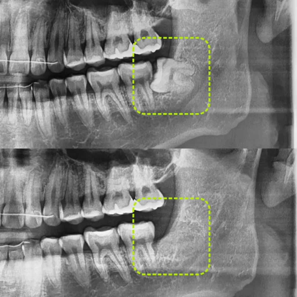
발치 전과 후의 파노라마를 비교해 보면, 신경관 가까이 자리하던 사랑니가 제거되어 정리된 것을 확인할 수 있습니다. 이렇게 신경과 밀접한 사랑니도 정확한 진단과 신중한 접근을 통해 발치가 가능한 경우가 있습니다.
사랑니 통증, 어떤 경우에 생기나요?
앞서 말씀드린 것처럼 사랑니 통증은 여러 원인으로 나타날 수 있습니다. 사랑니가 비스듬하게 나거나 일부만 맹출해 위생관리가 어려운 경우, 주변 잇몸에 염증(사랑니 주위염)이 반복되는 경우, 앞 어금니를 밀거나 짓눌러 손상을 주는 경우, 사랑니 자체에 충치가 진행된 경우 등이 대표적입니다.
이런 상태를 방치하면 통증뿐 아니라 앞 어금니 손상, 낭종 등 다른 문제로 이어질 수 있습니다. 따라서 통증이나 염증이 반복된다면 미루지 말고 검진을 받아보시는 것이 좋습니다.
신경과 가까운 사랑니, 무조건 위험하기만 한가요?
신경과 가까운 사랑니는 신중한 접근이 필요한 것은 맞지만, 무조건 발치가 불가능하다는 의미는 아닙니다. 이 사례처럼 3D CT로 신경과 뿌리의 관계를 정확히 파악하고, 최소 침습 원칙에 따라 조심스럽게 진행하면 발치가 가능한 경우가 있습니다.
반대로 신경과의 관계나 뿌리 형태에 따라 발치 난이도가 매우 높거나 특별한 주의가 필요한 경우도 있습니다. 따라서 신경과 가까운 사랑니는 반드시 영상검사를 통해 개별적으로 판단하는 것이 중요합니다.
사랑니, 시기를 놓치지 않는 것이 좋습니다
사랑니는 비교적 젊은 나이에 발치하는 편이 회복 속도가 빠르고 부담이 적은 경향이 있습니다. 시간이 지날수록 사랑니와 뼈의 유착이 진행되어 발치가 까다로워질 수 있고, 신경과 가까운 경우에는 더욱 신중한 접근이 필요해질 수 있습니다.
물론 모든 사랑니를 무조건 발치해야 하는 것은 아닙니다. 올곧게 자라 관리가 잘 되는 사랑니는 보존할 수 있으며, 발치 여부와 시기·방법은 개인의 사랑니 위치와 신경과의 관계에 따라 달라집니다. 중요한 것은 스스로 판단하기보다 검진과 영상검사를 통해 정확한 상태를 확인하는 것입니다.
자주 묻는 질문
Q. 사랑니 발치에 왜 CT까지 찍나요?
일반 파노라마는 2차원 영상이라 사랑니 뿌리와 신경관의 실제 거리·방향을 정확히 파악하기 어렵습니다. 이 사례처럼 신경과 가까운 경우에는 3차원 CT로 신경 위치를 미리 확인해야 보다 안전한 발치 계획을 세울 수 있습니다.
Q. 신경에 닿아 있는 사랑니도 뽑을 수 있나요?
신경과 가까울수록 신중한 접근이 필요하지만, 사전 CT 분석으로 위치를 파악하고 최소 침습으로 진행하면 발치가 가능한 경우가 있습니다. 다만 뿌리 형태와 신경 관계에 따라 난이도가 다르므로 정밀 진단이 중요합니다.
Q. 사랑니 발치 후 붓기와 통증이 심한가요?
과도한 절개와 조직 절제가 붓기·통증의 주요 원인입니다. 작게 절개하고 최소한으로 제거하면 붓기와 통증을 줄이는 데 도움이 될 수 있습니다. 다만 회복 양상은 개인차가 있습니다.
Q. 사랑니는 아프지 않으면 그냥 둬도 되나요?
올곧게 자라 관리가 잘 되면 보존할 수 있지만, 비스듬하거나 매복되어 있거나 앞 치아에 영향을 주는 경우에는 통증이 없어도 미리 검토하는 편이 좋은 경우가 많습니다. 증상 유무와 별개로 검진으로 확인하시길 권해 드립니다.
Q. 부분 매복된 사랑니는 왜 더 조심해야 하나요?
부분 매복 사랑니는 충치와 치주질환 발생 확률이 높아 치아 표면이 부식되어 있을 수 있습니다. 발치 시 조각이 으스러지지 않도록 신중하게 제거해야 하므로 더 조심스러운 접근이 필요합니다.
English Summary
Safely Extracting a Wisdom Tooth Whose Root Touched the Nerve Canal, Using 3D CT
Pain from a wisdom tooth can arise from several causes: an abnormal eruption pattern, inflammation of the surrounding gum, pressure against the second molar in front, or extensive decay in the tooth itself. Whatever the cause, when pain or inflammation appears, the best course is to visit a nearby dental clinic promptly for an examination. This case describes a wisdom tooth whose root tip appeared to touch the inferior alveolar nerve canal, and how it was extracted safely by first confirming the position of the nerve with a three-dimensional CT scan.
On the radiograph in this case, the root tip of the wisdom tooth appeared to be in contact with the nerve canal. The inferior alveolar nerve governs sensation in the lower lip and the area around the chin, so when the wisdom tooth root and the nerve canal lie this close together, extraction must be carried out with particular care, because forcing an approach near the nerve can lead to significant complications. For this reason, accurately grasping the relationship between the tooth root and the nerve in advance is the starting point for a safe extraction.
To extract safely, it is best to determine the exact positions of the nerve and the wisdom tooth root through three-dimensional CT imaging, because a standard panoramic image is two-dimensional and has limits in showing the true depth and the relationship to the nerve. In this case, the CT images were used to review the relationship between the wisdom tooth root and the nerve canal across multiple cross-sections.
The distance was indeed close, but the prior analysis indicated that extraction could be carried out. When a tooth is this closely related to the nerve, confirming the situation beforehand with CT is especially important.
To extract the wisdom tooth without severe swelling or pain afterward, minimizing tissue trauma is important; making a small incision and removing as little tooth structure as necessary results in much less post-operative pain. In addition, when a wisdom tooth is partially impacted, the likelihood of decay and periodontal disease is higher, so the tooth surface may well be eroded, and care must be taken to remove it in sections without crumbling.
Being close to the nerve, an unhurried and careful approach is the key. After the extraction, the follow-up image showed the wisdom tooth removed cleanly, without residual tissue, reflecting careful work guided by the prior CT analysis and the principle of minimal trauma. Comparing the panoramic images before and after, the wisdom tooth that had sat close to the nerve canal was cleared, illustrating that even a wisdom tooth closely related to the nerve can sometimes be extracted through accurate diagnosis and a careful approach.
Being close to the nerve calls for a cautious approach but does not automatically mean extraction is impossible; conversely, depending on the relationship to the nerve and the shape of the roots, some cases are of very high difficulty or require special caution, so a nerve-adjacent wisdom tooth should always be assessed individually through imaging. It is worth explaining why the CT step is so valuable in a case like this.
A panoramic film flattens the anatomy into a single plane, so a root and the nerve canal can appear to overlap even when a thin layer of bone still separates them, or they can look separated when they are in fact touching. A three-dimensional scan shows the same region slice by slice, revealing whether the nerve runs to the inner or outer side of the root and how much bone lies between them, and this is exactly the information that lets the surgeon plan how to section the tooth and in which direction to deliver each fragment so that pressure is directed away from the nerve rather than toward it.
More broadly, extracting a wisdom tooth at a relatively younger age tends to allow faster recovery with less burden, since over time the tooth and bone become more firmly fused and removal grows more difficult, and cases near the nerve may require even greater care. Not every wisdom tooth must be removed, however; a tooth that has erupted uprightly and is well maintained can be preserved, and whether, when, and how to extract depends on each person’s tooth position and nerve relationship. What matters most is not to judge on one’s own but to confirm the actual condition through examination and imaging, since individual anatomy and outcomes differ from person to person.
마치며
신경관에 근접한 사랑니는 발치 난이도가 높아 더욱 신중한 접근이 필요합니다. 이 사례처럼 사전 3D CT로 신경 위치를 정확히 확인하고, 최소 침습 원칙에 따라 신중하게 진행하면 붓기와 통증을 줄이면서 안전한 발치를 지향할 수 있습니다.
다만 사랑니와 신경의 관계, 뿌리 형태는 개인마다 다르므로 정확한 판단은 영상검사를 통해 이루어져야 합니다. 강동구, 명일동에서 사랑니 통증이나 신경과 가까운 사랑니 발치를 고민하고 계신다면 무리하지 마시고, 진료를 통해 본인의 사랑니 상태를 정확히 확인해 보시길 권해 드립니다. 임플란트 등 치과 치료 전반이 궁금하시다면 아래 글도 함께 참고해 보시면 도움이 되실 것입니다.
지금까지 서울더자연치과 치과의사 김민수였습니다.
※ 본 포스팅은 의료법 제56조 제1항의 의료광고 규정을 준수하여 작성되었으며, 실제 내원 환자분의 동의하에 공개된 사진입니다. 개인에 따라 진료 및 치료 방법, 효과와 부작용이 다르게 나타날 수 있으며, 진료 전 전문 의료진과의 충분한 상담을 권해 드립니다.