
신경관에 근접한 고난도 매복 사랑니, 3D CT로 안전하게 발치
신경관과의 거리가 1mm도 채 되지 않던 깊이 매복된 사랑니 사례입니다. 왜 고난도로 판단하는지, 3D CT와 최소 절개로 어떻게 안전을 우선해 발치했는지 사진과 함께 설명드립니다.
안녕하세요, 명일동 강동구 매복 사랑니 발치 전문 치과 서울더자연치과 치과의사 김민수입니다. 사랑니는 10대 후반에서 20대 초반에 맹출하는 세 번째 어금니로, 예쁜 이름과 달리 여러 문제를 일으키는 경우가 많습니다. 통증에서 끝나지 않고 고름이나 낭종, 음식물로 인한 충치와 치주질환, 구취까지 다양한 문제의 원인이 될 수 있습니다.
특히 사랑니는 바로 앞의 두 번째 어금니까지 손상시키는 경우가 많습니다. 올곧게 자라 관리가 잘 된다면 보존해도 괜찮지만, 잇몸 속에 묻혀 있거나 누워 있는 경우, 혹은 다른 치아에 좋지 않은 영향을 끼칠 때는 가급적 빠르게 발치를 검토하는 것이 좋습니다. 오늘은 그중에서도 신경관에 근접해 고난도로 판단된 매복 사랑니 사례를 예시 사진과 함께 정리해 드리겠습니다.
왜 고난도 케이스로 판단했나요
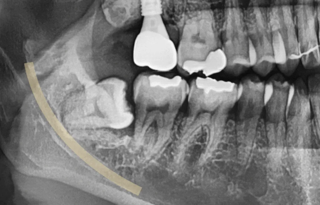
⬇️[01_초진_파노라마_신경관_근접]
내원 당시 파노라마를 관찰해 보면, 사랑니가 잇몸 속에 깊이 묻혀 있는 것을 확인할 수 있습니다. 표시해 둔 신경관과 단순히 가까이 위치한 정도가 아니라, 겹쳐 보일 정도로 인접해 있어 고난도 케이스로 판단되었습니다.
⬇️[02_치석_충치_근접_엑스레이]
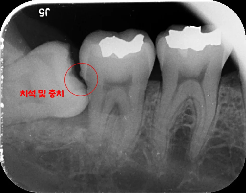
이와 더불어 앞 어금니 사이에 생긴 빈 공간에 음식물이 자주 끼면서 염증과 약간의 충치, 그리고 치석까지 함께 관찰되었습니다. 이런 상태가 지속되면 사랑니 자체뿐 아니라 앞 어금니 건강에도 영향을 줄 수 있어, 가급적 빠르게 정리하는 것이 앞으로의 구강 건강에 도움이 될 것으로 판단했습니다.
왜 3D CT 촬영이 필요한가요
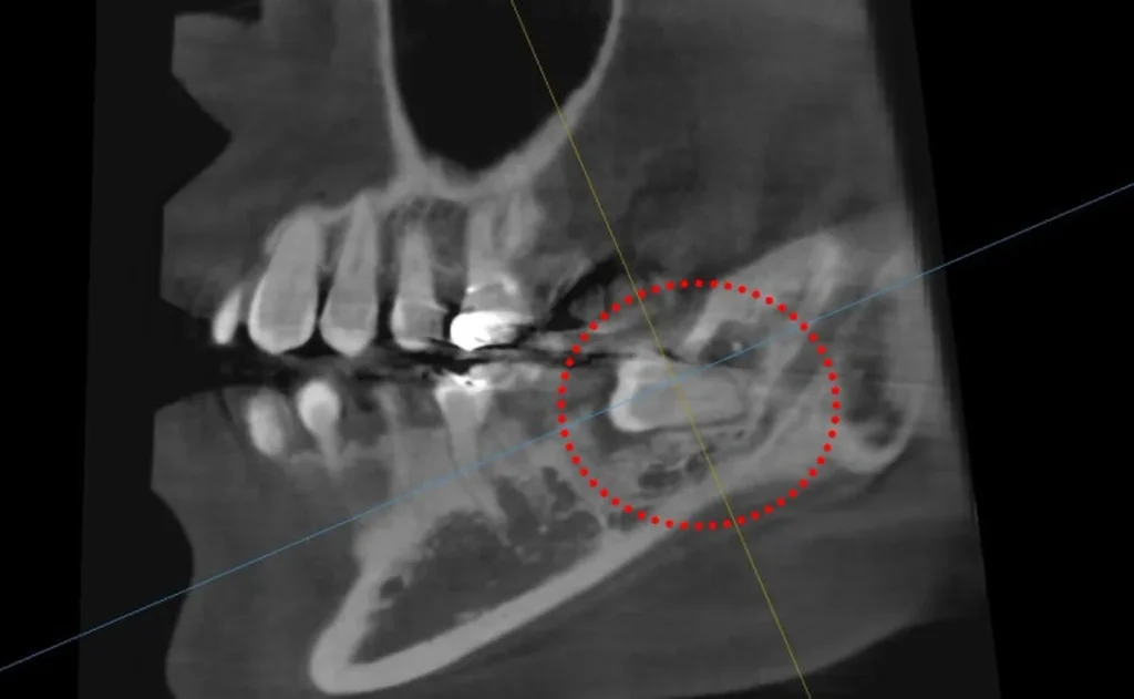
이렇게 신경관과 밀접하게 위치해 있을 때는 발치를 진행하기에 앞서 3차원 CT 촬영을 통해 신경의 위치를 정확히 확인해야 합니다. 일반 파노라마는 2차원 영상이라 실제 깊이와 신경과의 관계를 파악하는 데 한계가 있기 때문입니다.
상기 CT 촬영본을 보면 사랑니 뿌리와 신경관 사이의 거리가 약 1mm도 채 되지 않는 것으로 확인됩니다. 하치조신경은 아랫입술과 턱 주변의 감각을 담당하는 중요한 신경으로, 이 부위와의 관계를 미리 정밀하게 파악하는 것이 안전한 발치의 출발점이 됩니다. 이 사례처럼 신경과의 거리가 매우 가까운 경우에는 CT를 통한 사전 확인이 특히 중요합니다.
최소 절개로 어떻게 발치를 진행하나요
사랑니 발치 시 과도한 골삭제와 잇몸 절개는 발치 후 붓기와 통증의 원인이 됩니다. 따라서 이 사례에서는 꼭 필요한 부위만 최소한으로 절개하고, 안에 있는 치아머리 부분을 조각내어 제거하는 방식으로 접근했습니다. 이렇게 하면 붓기가 적고 통증도 최소화하는 데 도움이 될 수 있습니다.
앞서 계획한 대로 매복된 사랑니를 깨끗하게 발치하기 위해서는 조심스러운 접근이 필요합니다. 이는 신경과의 거리가 가깝기 때문이며, 치아 주변으로는 셀 수 없이 많은 혈관과 신경 등 조직이 존재하기 때문에 무엇보다 안전을 최우선으로 진행하는 것이 좋겠습니다.
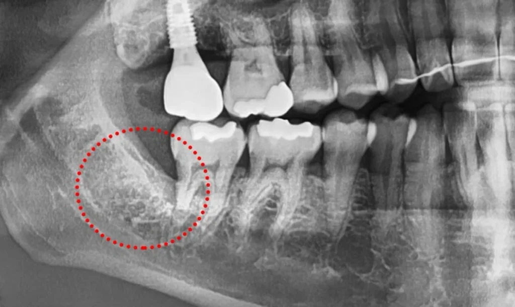
발치 후 예시 사진을 관찰해 보면 잔존 조직 없이 비교적 깨끗하게 제거된 것을 확인할 수 있습니다. 신경관에 근접한 고난도 케이스였던 만큼, 사전 CT 분석과 최소 절개 원칙에 따라 신중하게 진행한 결과입니다.
이런 경우라면 검진을 미루지 마세요
아직 사랑니를 뽑지 않고 계시거나, 원인 모를 통증이나 구취, 불편함이 생긴 분들은 가급적 빠르게 검진을 받아보시는 것이 중요합니다. 특히 이번 사례처럼 신경관에 근접한 매복 사랑니는 시간이 지날수록 발치 난이도와 부담이 커질 수 있습니다.
물론 모든 사랑니를 무조건 발치해야 하는 것은 아닙니다. 올곧게 자라 관리가 잘 되는 사랑니는 보존할 수 있으며, 발치 여부와 시기, 방법은 개인의 사랑니 위치와 신경과의 관계에 따라 달라집니다. 중요한 것은 스스로 판단하기보다 영상검사를 통해 정확한 상태를 확인하는 것입니다.
자주 묻는 질문
Q. 사랑니 발치에 왜 CT까지 찍나요?
일반 파노라마는 2차원 영상이라 사랑니 뿌리와 신경관의 실제 거리·방향을 정확히 파악하기 어렵습니다. 이 사례처럼 신경과 가까운 경우에는 3차원 CT로 신경 위치를 미리 확인해야 보다 안전한 발치 계획을 세울 수 있습니다.
Q. 신경에 가까운 사랑니는 위험하지 않나요?
신경과 가까울수록 신중한 접근이 필요한 것은 맞습니다. 그래서 사전 CT 분석으로 신경 위치를 파악하고, 최소 절개와 조심스러운 접근으로 안전을 우선해 진행합니다. 상태에 따라 주의사항이 다르므로 정밀 진단이 중요합니다.
Q. 발치 후 붓기와 통증이 많이 심한가요?
과도한 골삭제와 잇몸 절개가 붓기·통증의 주요 원인입니다. 꼭 필요한 부위만 최소 절개하고 치아를 조각내어 제거하면 붓기와 통증을 줄이는 데 도움이 될 수 있습니다. 다만 회복 양상은 개인차가 있습니다.
Q. 사랑니는 아프지 않으면 그냥 둬도 되나요?
올곧게 자라 관리가 잘 되면 보존할 수 있지만, 잇몸 속에 묻히거나 누워 있거나 앞 치아에 영향을 주는 경우에는 통증이 없어도 미리 검토하는 편이 좋은 경우가 많습니다. 증상 유무와 별개로 검진을 통해 확인하시길 권해 드립니다.
Q. 매복 사랑니가 앞 어금니에도 영향을 주나요?
그렇습니다. 이 사례처럼 사랑니와 앞 어금니 사이 빈 공간에 음식물이 끼면 염증·충치·치석이 생길 수 있고, 평생 사용해야 할 두 번째 어금니까지 손상될 수 있습니다. 이 때문에 문제가 커지기 전 발치를 검토하는 경우가 많습니다.
English Summary
Safely Extracting a High-Difficulty Impacted Wisdom Tooth Close to the Nerve Using 3D CT
Wisdom teeth are the third molars, usually erupting in the late teens or early twenties. Despite the affectionate name they carry in Korean, they frequently cause trouble. Problems do not stop at pain; they can lead to pus or cysts, decay and periodontal disease from trapped food, and bad breath, and they often damage the second molar directly in front of them, a tooth that has to last a lifetime. When a wisdom tooth erupts uprightly and can be kept clean, it may be preserved, but when it is buried in the gum, lying on its side, or harming the neighboring teeth, earlier extraction is generally preferable. This case describes one such tooth that was judged high-difficulty because of its closeness to the nerve.
On the initial panoramic radiograph, the wisdom tooth was buried deep within the gum. It was not merely near the marked nerve canal; it appeared to overlap with it, which is why the case was classified as high-difficulty. Alongside this, the empty space that had formed between the wisdom tooth and the adjacent molar had been trapping food, and inflammation, slight decay, and calculus were all visible. Left unaddressed, such a situation can affect not only the wisdom tooth itself but also the health of the second molar in front, so clearing it sooner was judged better for long-term oral health.
When a wisdom tooth sits this close to the nerve canal, a three-dimensional CT scan is taken before extraction to confirm the exact position of the nerve. A standard panoramic image is two-dimensional, so it has limits in showing the true depth and the relationship to the nerve. On the CT images in this case, the distance between the tooth root and the nerve canal measured less than about one millimeter. The inferior alveolar nerve governs sensation in the lower lip and the area around the chin, so understanding its relationship to the tooth in advance is the starting point of a safe extraction. When the nerve is this close, prior confirmation with CT is especially important.
Excessive bone removal and gum incision during a wisdom tooth extraction are the main causes of post-operative swelling and pain. For this reason, the approach here was to incise only the strictly necessary area and to section the crown of the tooth inside before removing it, which helps keep swelling and pain to a minimum. Removing a deeply impacted tooth cleanly, as planned, requires a careful approach precisely because the nerve is nearby. Since countless blood vessels, nerves, and other tissues surround the tooth, prioritizing safety above all else is the guiding principle.
After the extraction, the follow-up image showed that the tooth had been removed cleanly, without residual tissue. Given that this was a high-difficulty case close to the nerve, this outcome reflected careful work guided by the prior CT analysis and the principle of minimal incision. The broader message is a practical one: those who have not yet had a troublesome wisdom tooth removed, or who have developed unexplained pain, bad breath, or discomfort, are encouraged to seek an examination sooner rather than later, because a nerve-adjacent impacted tooth tends to become more difficult to remove as time passes.
None of this means every wisdom tooth must be extracted. A tooth that has erupted uprightly and is well maintained can be preserved, and the decision to extract, along with its timing and method, depends on each person’s tooth position and its relationship to the nerve. What matters most is not to judge on one’s own but to confirm the actual condition through imaging. Because individual anatomy differs so much, and because outcomes and recovery vary from person to person, an in-person examination and consultation are recommended before any decision is made about a wisdom tooth of this kind.
It is also worth explaining why the minimal-incision approach matters so much in a case like this. The more bone is removed and the wider the gum is opened, the more the surrounding tissue is disturbed, and that disturbance is what the body responds to with swelling and soreness in the days that follow. By opening only what is needed and dividing the buried crown into smaller pieces, the tooth can often be delivered through a smaller window, which tends to shorten the recovery and reduce discomfort. This is not a guarantee of a painless recovery, since every patient heals differently, but it reflects a deliberate effort to keep the procedure as gentle as the anatomy allows while still removing the tooth completely and confirming, on a follow-up image, that no fragments remain behind near the nerve.
마치며
신경관에 근접한 매복 사랑니는 발치 난이도가 높아 더욱 신중한 접근이 필요합니다. 이 사례처럼 사전 3D CT로 신경 위치를 정확히 확인하고, 최소 절개 원칙에 따라 안전을 우선하면 붓기와 통증을 줄이면서 깨끗한 발치를 지향할 수 있습니다.
우리의 치아 건강은 지킬 수 있을 때 지키는 것이 중요합니다. 강동구, 명일동에서 매복 사랑니나 원인 모를 통증·구취·불편함으로 고민하고 계신다면 무리하지 마시고, 진료를 통해 본인의 사랑니 상태를 정확히 확인해 보시길 권해 드립니다. 임플란트 등 치과 치료 전반이 궁금하시다면 아래 글도 함께 참고해 보시면 도움이 되실 것입니다.
지금까지 서울더자연치과 치과의사 김민수였습니다.
※ 본 포스팅은 의료법 제56조 제1항의 의료광고 규정을 준수하여 작성되었으며, 실제 내원 환자분의 동의하에 공개된 사진입니다. 개인에 따라 진료 및 치료 방법, 효과와 부작용이 다르게 나타날 수 있으며, 진료 전 전문 의료진과의 충분한 상담을 권해 드립니다.