
수면치료로 매복 사랑니 발치와 어금니 임플란트까지
매복 사랑니(38번) 때문에 앞 어금니(37번)까지 손상된 사례입니다. 발치에 대한 부담을 줄이기 위해 의식하진정법(수면치료)을 적용해 발치와 임플란트 식립을 진행한 과정을 사진과 함께 정리해 드립니다.
안녕하세요, 명일동 강동구에 위치한 서울더자연치과 치과의사 김민수입니다. 사랑니는 대개 10대 후반에서 20대 초반에 맹출하는 세 번째 어금니로, 구강에서 가장 늦게 자라기 때문에 잇몸 맨 뒤쪽의 좁은 틈을 비집고 나오는 경우가 많습니다. 그러다 보니 옆으로 눕거나 잇몸 속에 묻히는 등 변형된 형태로 자리 잡는 경우가 적지 않습니다.
문제는 이렇게 매복된 사랑니가 본인의 문제로 끝나지 않고, 바로 앞 어금니까지 함께 망가뜨릴 수 있다는 점입니다. 오늘 소개해 드릴 사례가 바로 그런 경우로, 발치에 대한 두려움이 크셨던 분이라 의식하진정법(수면치료)을 적용해 매복 사랑니 발치와 어금니 임플란트를 함께 진행했습니다.
초기 상태는 어땠나요

내원 당시 구내 사진을 보면, 잇몸 속에 자리한 세 번째 어금니(사랑니, 38번) 때문에 바로 앞의 두 번째 어금니(37번)가 심하게 손상된 것으로 보입니다. 매복 사랑니와 앞 어금니 사이는 칫솔이 닿지 않아 음식물과 세균이 계속 머물기 쉬운 부위이기 때문입니다.
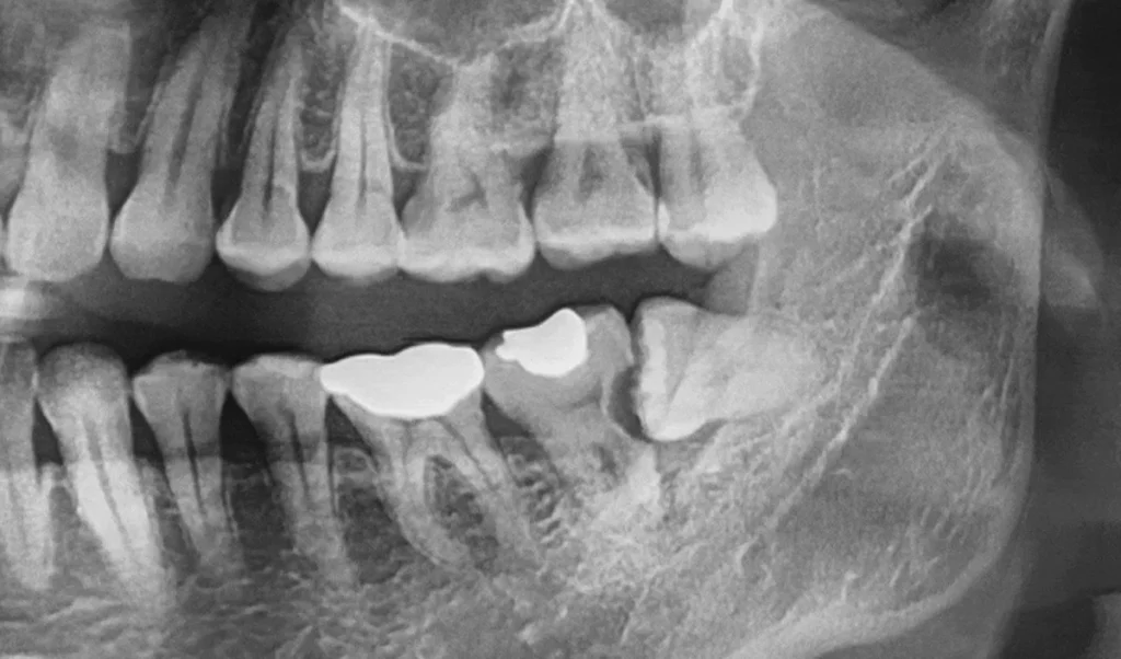
구내 사진만으로는 잘 드러나지 않지만, 파노라마 엑스레이를 확인하면 상황이 훨씬 분명해집니다. 원심면 치근-치관 사이에 광범위한 우식(충치)이 진행되어 주변 치조골 소실까지 나타나 있는 것으로 보입니다.
이 사례에서는 37번과 38번 모두 발치가 필요한 상황으로 판단되었습니다. 따라서 두 곳을 정리한 뒤, 기능을 담당하던 어금니인 37번 자리에는 임플란트 식립이 필요하겠다고 보았습니다.
왜 수면치료(의식하진정법)를 함께 고려했나요
매복 사랑니 발치와 임플란트 식립을 한 번에 계획하면 시술 시간이 다소 길어질 수 있고, 발치 자체에 대한 두려움이 크신 분들은 진료 자체를 미루다가 상태가 더 나빠지는 경우가 있습니다. 이 사례의 환자분도 치과 치료에 대한 부담이 크셨던 상황이었습니다.
이런 경우 의식하진정법(수면치료)이 도움이 될 수 있습니다. 의식하진정법은 완전히 잠드는 전신마취와 달리, 진정제를 이용해 긴장과 불안을 낮추고 몸의 힘을 빼 편안한 상태에서 치료를 받도록 돕는 방식입니다. 자발 호흡은 유지되며, 시술 중의 긴장과 불안, 시간에 대한 부담을 줄이는 데 목적이 있습니다.
다만 수면치료가 모든 분께 항상 필요한 것은 아니며, 전신 건강 상태와 복용 약물, 시술 범위 등을 함께 고려해 적용 여부를 판단하는 것이 좋겠습니다. 진정 상태에서도 활력징후를 지속적으로 관찰하며 안전에 유의해 진행하게 됩니다.
발치부터 임플란트, 최종 보철까지 어떻게 진행되나요
진행 과정을 순서대로 정리해 드리겠습니다.
1. 매복 사랑니와 손상된 어금니 발치
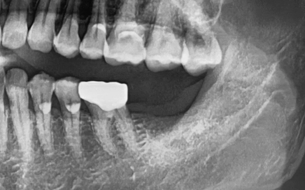
사랑니가 잇몸 속에 묻혀 있는 형태로 보였기 때문에, 최소한의 절개를 통해 필요한 경우 치아를 조각내어 조심스럽게 빼내는 방식으로 진행했습니다. 앞 어금니(37번)는 심한 충치로 표면이 부식되어 있어, 치아 조각이 떨어지거나 주변 조직에 영향을 주지 않도록 특히 조심스럽게 발치를 진행했습니다.
발치 후에는 엑스레이 촬영을 통해 잔존 조직이 남아 있지 않은지 점검합니다. 예시 사진을 보면 잔존 조직 없이 비교적 깔끔하게 발치가 마무리된 것으로 보입니다. 수면치료를 병행한 덕분에 발치 과정에 대한 부담을 상당히 덜 수 있었습니다.
2. 임플란트 식립
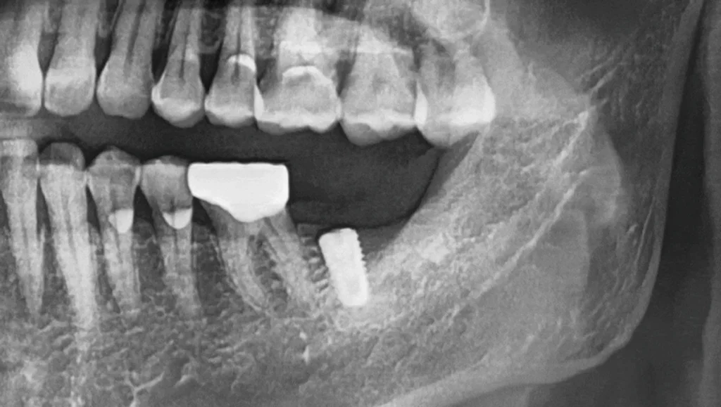
발치 후 잇몸이 어느 정도 회복되면 임플란트를 식립하는 과정을 진행할 수 있습니다. 이때 신경과 주변 인접치, 그리고 대합치(맞물리는 치아)와의 관계를 함께 고려하여 최적의 식립 위치를 결정하는 것이 중요합니다.
계획한 위치에 픽스처를 식립하면, 파노라마에서 안정적으로 자리 잡은 픽스처를 확인할 수 있습니다. 하악 어금니 부위는 신경관과의 거리를 신중하게 살펴야 하는 부위이기에, 식립 전 영상 분석과 위치 계획이 특히 중요합니다.
3. 크라운(최종 보철) 제작
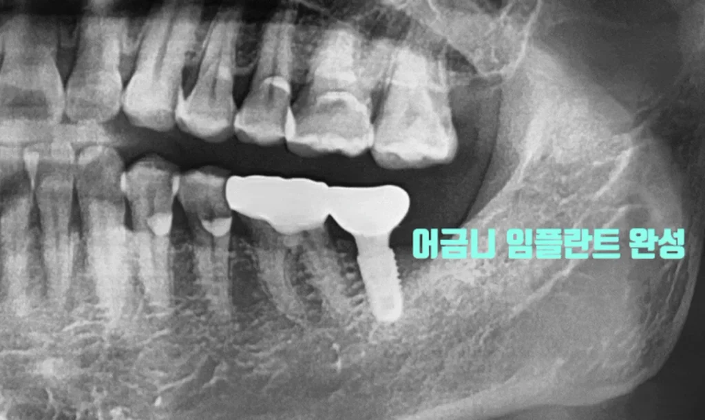
픽스처가 단단하게 자리를 잡을 수 있도록 약 2개월간 골 유착 시간을 둡니다. 이후 ISQ 수치 측정을 통해 고정력을 확인한 뒤 최종 크라운을 제작해 올리게 됩니다. 다만 골 유착 기간과 고정력은 개인의 뼈 상태와 전신 조건에 따라 달라질 수 있습니다.
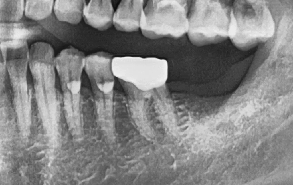
최종 보철까지 마무리되면, 매복 사랑니 때문에 손상되었던 어금니 자리가 임플란트로 다시 기능을 되찾은 것을 확인할 수 있습니다. 이렇게 매복 사랑니로 인해 앞 치아까지 손상된 경우에도, 발치와 임플란트를 계획적으로 연결해 어금니 기능을 재건할 수 있습니다.
사랑니, 왜 시기를 놓치지 않는 것이 좋을까요
이 사례가 보여주듯, 매복 사랑니는 방치하면 본인뿐 아니라 앞 어금니까지 함께 손상시킬 수 있습니다. 옆으로 누운 사랑니와 앞니 사이에는 양치가 불가능한 틈이 생기고, 그 자리에 충치나 낭종 등이 생겨 문제가 커지기 전에 미리 정리하는 편이 좋은 경우가 많습니다.
또한 비교적 젊은 나이에 발치하는 편이 회복 속도가 빠르고 부담이 적은 경향이 있습니다. 반대로 40대 이상이 되면 사랑니와 치조골의 골유착이 진행되어 발치가 까다로워지고 회복에 더 부담이 될 수 있는 것으로 보입니다. 물론 발치 시기와 방법은 개인의 상태에 따라 다르므로, 정확한 판단은 검진을 통해 확인하는 것이 좋겠습니다.
자주 묻는 질문
Q. 수면치료(의식하진정법)를 하면 완전히 잠드나요?
전신마취처럼 완전히 잠드는 것과는 다릅니다. 진정제를 이용해 긴장과 불안을 낮추고 편안한 상태를 유도하는 방식으로, 자발 호흡은 유지됩니다. 시술에 대한 부담과 긴장을 줄이는 데 목적이 있습니다.
Q. 매복 사랑니 때문에 앞니까지 꼭 빼야 하나요?
앞 어금니의 손상 정도에 따라 다릅니다. 이 사례처럼 충치가 광범위하게 진행되어 살리기 어려운 경우에는 발치 후 임플란트 등으로 재건하기도 하며, 손상이 크지 않다면 치료 후 보존하는 경우도 있습니다. 정밀 진단이 우선입니다.
Q. 발치와 임플란트를 같은 날 하나요?
상태에 따라 다릅니다. 발치 후 잇몸과 뼈의 회복을 확인한 뒤 임플란트를 식립하는 경우가 많으며, 이 사례에서도 발치 후 회복을 거쳐 식립을 진행했습니다. 순서와 시점은 진단을 통해 결정됩니다.
Q. 임플란트 후 바로 크라운을 씌우나요?
그렇지 않습니다. 픽스처가 뼈와 유착되고 고정력이 확인되어야 최종 크라운을 올릴 수 있습니다. 이 사례에서는 약 2개월의 골 유착 기간을 두고 ISQ 수치로 고정력을 확인한 뒤 보철을 제작했으며, 기간은 개인차가 있습니다.
Q. 사랑니는 아프지 않으면 그냥 둬도 되나요?
매복 사랑니는 통증이 없어도 앞 치아에 충치나 낭종 등을 유발할 수 있습니다. 특히 옆으로 누운 형태라면 문제가 생기기 전에 미리 검토하는 편이 좋은 경우가 많으므로, 증상 유무와 별개로 검진을 통해 상태를 확인하시길 권해 드립니다.
English Summary
Impacted Wisdom Tooth Extraction and Molar Implant Under Conscious Sedation
Wisdom teeth are the third molars, usually erupting in the late teens or early twenties. Because they are the last teeth to develop and must push through the narrow space at the very back of the jaw, they frequently end up lying on their side or remaining buried within the gum.
A problem with an impacted wisdom tooth does not always stay confined to that tooth; it can damage the neighboring second molar as well. This case is exactly that situation, and because the patient felt considerable anxiety about extraction, conscious sedation was used so that the wisdom tooth removal and the implant placement could be carried out comfortably.
At the initial visit, the intraoral photograph suggested that the buried third molar had contributed to severe damage in the second molar directly in front of it. The gap between an impacted wisdom tooth and the adjacent tooth cannot be reached by a toothbrush, so food debris and bacteria tend to accumulate there. The intraoral view alone did not reveal the full extent of the problem, but the panoramic radiograph made it clear: extensive decay had developed between the root and crown on the distal surface, and loss of the surrounding alveolar bone was also evident. Both the second molar and the wisdom tooth were judged to require extraction, and the second molar site was planned for implant restoration afterward.
Conscious sedation was considered alongside the surgical plan because combining an impacted extraction with implant placement can lengthen the appointment, and patients with a strong fear of extraction sometimes delay treatment until their condition worsens. Unlike general anesthesia, conscious sedation uses medication to reduce tension and anxiety while the patient breathes on their own, staying relaxed throughout the procedure.
It is not always necessary for everyone; general health, medications, and the scope of the procedure are all weighed when deciding whether to use it, and vital signs are monitored continuously for safety.
The procedure was carried out in sequence. Because the wisdom tooth appeared buried within the gum, a minimal incision was made and the tooth was sectioned and removed carefully where needed. The second molar in front, its surface eroded by severe decay, was extracted with particular care so that fragments would not break away or affect the surrounding tissue. A follow-up radiograph confirmed that the sites had been cleared without residual tissue. Because sedation was used, the burden of the extraction phase was considerably reduced for the patient.
Once the gum had healed to a suitable degree, the implant was placed. The optimal position was determined by considering the nerve, the adjacent teeth, and the opposing tooth, which is especially important in the lower molar region where the distance to the nerve canal must be assessed carefully in advance. After placement, the panoramic image showed the fixture stably positioned.
The site was then given about two months for osseointegration, after which the stability was checked using an ISQ measurement before the final crown was fabricated. Healing time and stability vary with each person’s bone condition and general health.
When the final restoration was completed, the molar site that had been compromised by the impacted wisdom tooth had regained its function through the implant. This case illustrates that even when an impacted wisdom tooth has damaged the tooth in front of it, a planned sequence connecting extraction and implant placement can restore molar function. It also highlights why timing matters: an impacted, sideways wisdom tooth creates a cleaning-inaccessible gap where decay and cysts can form, so addressing it before problems escalate is often preferable.
Extraction at a relatively younger age tends to allow faster recovery with less burden, whereas after the age of forty the wisdom tooth and bone may become more firmly fused, making removal more difficult. The appropriate timing and method differ from person to person and are best confirmed through an in-person examination rather than a fixed rule applied to everyone.
A further point worth emphasizing is the role of sedation in encouraging timely care. For patients whose fear has kept them from the dental chair, the discomfort they imagine is often what postpones treatment, and that delay is precisely what allows a single impacted tooth to compromise the tooth beside it. By lowering the anxiety associated with a longer combined procedure, conscious sedation can make it more feasible to address both the extraction and the reconstruction while the situation is still manageable.
The goal is never sedation for its own sake, but a treatment plan the patient can actually complete, carried out with continuous monitoring and appropriate caution.
마치며
매복 사랑니는 통증이 없더라도 앞 치아까지 손상시킬 수 있고, 방치할수록 발치와 이후 치료의 부담이 커질 수 있습니다. 이 사례처럼 앞 어금니가 이미 손상된 경우에도, 발치와 임플란트를 계획적으로 연결하고 필요에 따라 수면치료를 병행하면 부담을 줄이며 어금니 기능을 재건할 수 있습니다.
강동구, 명일동에서 매복 사랑니 발치나 발치에 대한 두려움, 어금니 임플란트를 고민하고 계신다면 무리하지 마시고 진료를 통해 본인의 구강 상태를 정확히 확인해 보시길 권해 드립니다. 임플란트 치료 전반이 궁금하시다면 아래 글도 함께 참고해 보시면 도움이 되실 것입니다.
지금까지 서울더자연치과 치과의사 김민수였습니다.
※ 본 포스팅은 의료법 제56조 제1항의 의료광고 규정을 준수하여 작성되었으며, 실제 내원 환자분의 동의하에 공개된 사진입니다. 개인에 따라 진료 및 치료 방법, 효과와 부작용이 다르게 나타날 수 있으며, 진료 전 전문 의료진과의 충분한 상담을 권해 드립니다.