
RE-ENDODONTIC TREATMENT
발치 대신 자연치아를 지키는 재신경치료(재근관치료)
안녕하세요, 재신경치료 전문 서울더자연치과 치과의사 김민수입니다. 오래전 신경치료를 받고 크라운(보철)을 씌운 어금니라도, 시간이 지나면 다시 문제가 생기는 경우가 있습니다. 이럴 때 무조건 발치하고 임플란트로 가는 것이 아니라, 이미 치료했던 신경관을 다시 열어 재치료하는 재신경치료(re-endo)로 자연치를 살릴 수 있는 경우가 적지 않습니다. 이번 글에서는 36번 어금니에 이차 충치와 뿌리 끝 염증이 관찰된 환자분의 실제 진행 과정을 정리해 보았습니다.
01
DIAGNOSIS
보철 아래 숨어 있던 이차 충치와 마진 들뜸
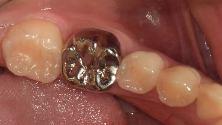
36번(좌측 첫 번째 큰 어금니)에 통증과 염증이 관찰되었습니다. 교합면을 확인해보면 이미 금속 보철(크라운)이 씌워져 있는 상태였습니다.
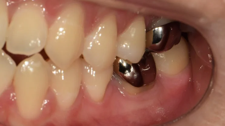
옆쪽 사진을 관찰해보니 보철의 마진(치아와 보철이 맞닿는 경계)이 들떠 있는 것을 확인할 수 있었습니다. 이차 충치가 의심되는 소견입니다. 이렇게 보철의 적합도가 떨어지는 경우에는, 대부분 외부의 이물질이나 세균이 내부로 침투하여 문제를 일으킬 수 있어 내부 상황이 좋지 않을 것으로 보입니다.
02
EXAMINATION
파노라마로 확인한 뿌리 끝 염증
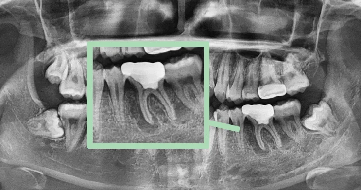
파노라마 촬영본을 관찰하며 감염이 어디까지 진행되었는지 확인해보았습니다. 치근(뿌리) 주변으로 검게 변해 있는 염증 소견이 관찰됩니다. 상황이 생각보다 좋지 않을 것으로 예상되는 상태로, 심하게 퍼진 염증으로 인해 치아가 흔들리는 것은 물론 심한 통증까지 느낄 수 있는 단계로 보입니다.
이러한 경우, 재신경치료(re-endo)를 진행하여 문제를 개선하는 것이 좋겠다고 판단됩니다.
03
TREATMENT PROCESS
자연치아를 살리는 재신경치료 과정
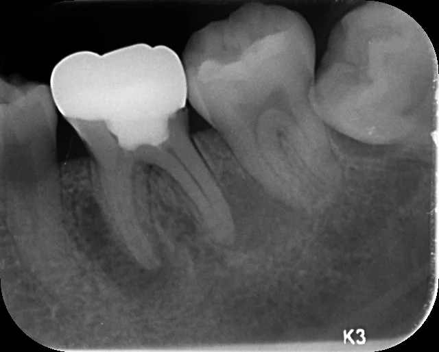
재신경치료를 통해 뿌리 끝 염증을 확실하게 제거하고, 근관 내부를 세척한 뒤 소독해줍니다. 재신경치료는 이미 치료를 받았던 곳을 다시 제거하고 소독·충전하기 때문에, 처음 하는 신경치료보다 더 어렵고 까다로운 과정입니다.
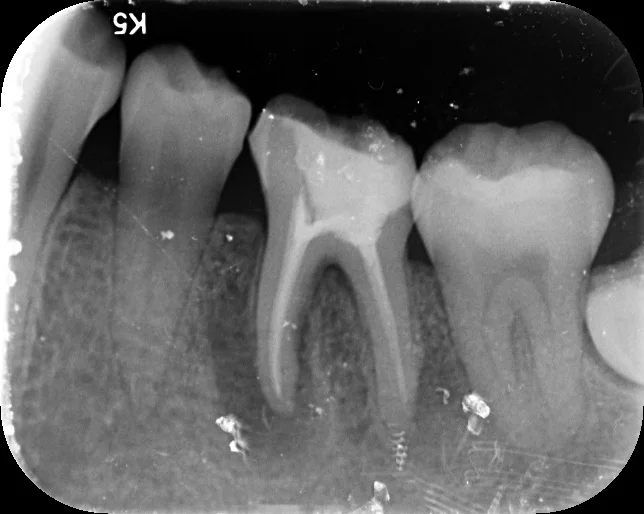
뿌리 끝 염증을 확실하게 치료한 다음, 근관을 충전하고 그 위에 크라운을 수복해줍니다. 감염된 조직을 남김없이 정리하는 것이 재치료 성공의 핵심이라고 보입니다.
최종 점검과 크라운 수복
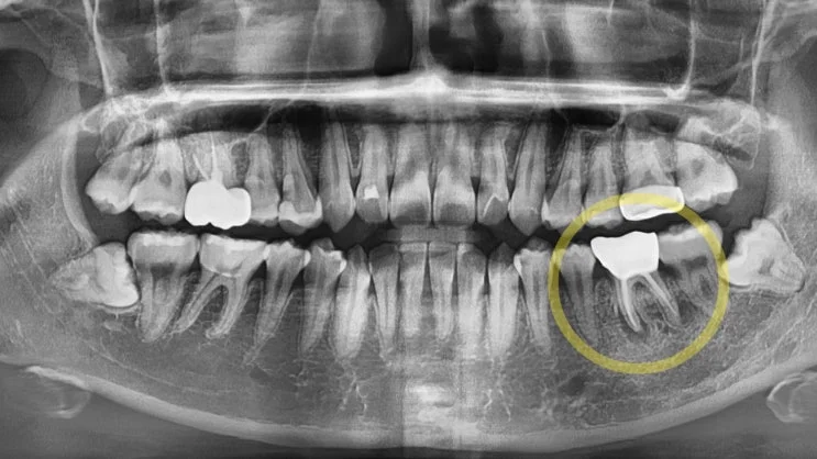
엑스레이를 통해 마지막으로 점검하면 과정이 마무리됩니다. 파노라마상 해당 부위에 지르코니아 크라운을 세팅한 것으로 보입니다. 근관 충전과 보철이 안정적으로 자리 잡은 모습입니다.
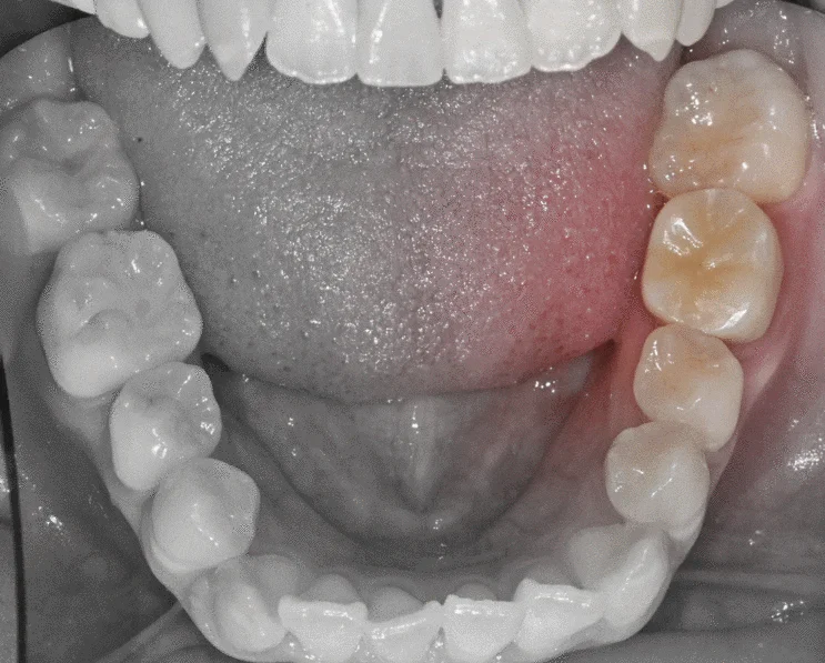
최종 교합면 사진입니다. 발치하지 않고 환자분 본인의 자연치를 그대로 유지하면서, 기능과 심미를 회복한 결과입니다. 긴 글 읽어주셔서 감사합니다. 김민수 대표원장이었습니다.
자주 묻는 질문 (FAQ)
Q. 신경치료 받고 크라운까지 씌운 치아인데, 또 아플 수 있나요?
네, 가능합니다. 신경치료 후 오랜 시간이 지나 보철 마진이 들뜨거나 이차 충치가 생기면 세균이 다시 내부로 침투해 뿌리 끝에 염증이 재발할 수 있습니다. 통증이나 잇몸 붓기가 있다면 방치하지 마시고 검진받아 보시는 것이 좋습니다.
Q. 재신경치료와 처음 하는 신경치료는 무엇이 다른가요?
재신경치료(re-endo)는 이전에 충전되어 있던 재료를 먼저 제거한 뒤 다시 소독·충전하는 과정입니다. 기존 치료 부위를 정리하고 다시 접근해야 하므로 일반 신경치료보다 시간과 정밀함이 더 요구되는 편입니다.
Q. 무조건 뽑고 임플란트 하는 것보다 재신경치료가 나은가요?
자연치아는 임플란트가 대신하기 어려운 고유의 감각과 기능을 가지고 있어, 살릴 수 있다면 자연치를 유지하는 것이 우선적으로 고려됩니다. 다만 염증 범위, 뿌리와 치아 잔존 상태에 따라 재신경치료의 예후가 달라지므로, 검진 후 개별적으로 판단하는 것이 좋겠습니다.
Q. 재신경치료는 아픈가요? 몇 번 정도 내원해야 하나요?
국소마취 하에 진행하며, 염증 정도와 근관 상태에 따라 내원 횟수는 달라질 수 있습니다. 근관 내부를 충분히 소독해야 하는 경우 여러 차례 나누어 진행하기도 합니다.
Q. 재신경치료 후에는 꼭 크라운을 해야 하나요?
신경치료를 받은 치아는 영양 공급이 줄어 상대적으로 약해지고 깨지기 쉬워집니다. 특히 어금니는 씹는 힘을 많이 받기 때문에, 치아를 감싸주는 크라운 수복이 권장되는 것이 일반적입니다.
English Summary – Re-Endodontic Treatment to Save a Natural Tooth (Case of Tooth #36)
Losing a natural tooth can be emotionally overwhelming, and at Seoul The Nature Dental Clinic in Gangdong-gu, our priority is to preserve a patient’s own teeth whenever it is clinically reasonable. This clinical record documents a case involving tooth #36, the first molar on the lower left, which presented with pain and signs of inflammation. Rather than moving directly to extraction and an implant, we evaluated whether re-endodontic treatment could save the tooth.
On examination of the occlusal surface, the tooth was already restored with a metal crown from a previous treatment. When we examined the lateral view, we could clearly see that the margin of the crown—the boundary where the restoration meets the natural tooth—was lifted and no longer sealing properly. A poorly fitting margin like this strongly suggests secondary caries. When the fit of a restoration deteriorates, external debris and bacteria can penetrate underneath, leading to problems developing deep inside the tooth where they are not immediately visible.
To determine how far the infection had spread, we reviewed the panoramic radiograph. Around the root of the tooth, a dark radiolucent change consistent with inflammation was observed. This indicated that the situation was more advanced than it might appear on the surface. When inflammation spreads extensively around the root, it can cause the tooth to become mobile and can also produce significant pain. Based on these findings, we judged that re-endodontic treatment (re-endo) would be the appropriate approach to resolve the problem and give the tooth a chance to be retained.
Re-endodontic treatment is more demanding than an initial root canal procedure. Because the tooth has already been treated once, the existing filling material inside the canals must first be carefully removed before the canals can be cleaned, disinfected, and re-filled. Through this process, the inflammation at the root tip is thoroughly removed, and the interior of the root canal system is irrigated and disinfected. Only after the infected tissue has been fully addressed can the tooth be reliably restored.
Once the root tip inflammation was managed, the canals were sealed and a crown was placed over the tooth to protect it. Teeth that have undergone root canal therapy tend to become more brittle because their internal blood and nutrient supply is reduced, and molars in particular bear heavy chewing forces. For this reason, covering the tooth with a crown is generally recommended. A follow-up X-ray was taken as a final check to confirm that the canal filling and restoration were stable and well positioned; the radiograph showed what appeared to be a zirconia crown seated on the tooth.
The final occlusal photograph shows the completed result, with the patient’s own natural tooth preserved and both function and esthetics restored, without the need for extraction. This case illustrates an important principle in dentistry: an implant, however excellent, cannot fully replicate the sensation and function of a natural tooth. When a tooth can be saved, retaining it is usually considered first. However, the prognosis of re-endodontic treatment depends on the extent of inflammation and the remaining structure of the root and crown, so the decision must always be made individually after a proper clinical and radiographic examination.
It is worth emphasizing why a lifted crown margin is such an important warning sign. A crown is meant to seal the underlying tooth completely, acting as a barrier against oral bacteria. When that seal breaks down—whether from long-term wear, recurrent decay at the edge, or a gap that develops over time—saliva and bacteria can seep into the space between the crown and the tooth. This creates an environment where secondary caries progresses silently, often without obvious symptoms until the infection reaches the pulp chamber or the root tip.
By the time a patient notices pain or swelling, the inflammation has frequently already extended into the surrounding bone, which is what the dark radiolucent area on the panoramic image represented in this case. Early detection through regular check-ups is therefore far more protective than waiting for discomfort to appear.
Aftercare also plays a meaningful role in the long-term success of a re-treated tooth. Following re-endodontic treatment and crown placement, patients are generally advised to avoid biting very hard foods directly on the restored molar for a period, to maintain thorough brushing and interdental cleaning around the crown margin, and to attend periodic recall visits so the tooth and surrounding bone can be monitored radiographically. Because the tooth no longer has a vital pulp, it will not signal problems through temperature sensitivity the way a healthy tooth would, which makes routine professional monitoring especially valuable.
If you are experiencing pain or gum swelling around a previously treated tooth in the Myeongil-dong or Gangdong-gu area, we recommend a professional evaluation before assuming that extraction is the only option. All clinical facts should be verified with your treating dentist prior to any procedure.
마치며
한 번 신경치료를 받은 치아라도, 상황에 따라 다시 살릴 수 있는 방법이 남아 있는 경우가 있습니다. 통증이나 잇몸 붓기를 오래 참기보다는, 정확한 검진을 통해 발치 전에 자연치를 지킬 수 있는지 먼저 확인해보시길 권해드립니다. 서울더자연치과는 명일동·강동구에서 환자분의 자연치를 최대한 보존하는 방향을 우선으로 진료하고 있습니다.
함께 보면 좋은 글
임플란트가 궁금하시다면 강동구 임플란트 완전 가이드 2026 글도 함께 참고해보시기 바랍니다.
※ 본 포스팅은 의료법 제56조 제1항을 준수하여 작성되었습니다. 실제 내원 환자분의 동의하에 공개된 사진이며, 동일한 환자분께 동일한 조건에서 촬영한 사진을 활용하였습니다. 다만 개인에 따라 진료 및 치료 방법이 다르게 적용될 수 있으며, 효과와 부작용이 다르게 나타날 수 있습니다. 진료 전 전문 의료진과의 충분한 상담을 권해드립니다.